What are NAMs?
Written by Gabby Vidaurre, Ph.D.
September 2025
Recently, the National Institutes of Health (NIH) announced several new initiatives aimed at reducing the use of animals in biomedical research and increasing the use of human-based research technologies, commonly referred to as NAMs, which stands for new approach methodologies or non-animal methods.1,2 Major reasons for this shift include the high failure rate of drugs once they reach human clinical trials and the growing body of literature documenting the problems with translating findings from animals to humans. Historically, drugs have often been developed and first tested in animals during preclinical trials; however, once they move into clinical trials, they frequently fail to improve the condition they were designed to treat and even cause serious side effects that were not observed in animals.3
So what exactly are NAMs?
NAMs use human cells, tissues, or clinical data to study human diseases. At SAO, we define NAMs as non-animal methods, meaning completely human. The term “new approach methodologies” is more often used in a toxicological context and sometimes includes techniques or data derived from animals. While using animal cells or data can reduce the use of live animals, it distracts from the point of these technologies: to generate findings that are applicable to humans.4 For example, using a mouse cell line to study Alzheimer’s disease may reduce suffering by avoiding the additional use of live mice, but what’s the point of using information from a species that doesn’t even get Alzheimer’s in the first place?
Because they rely on human-based materials and information, NAMs are more relevant to human biology and have already shown promise in better predicting how drugs will perform in clinical trials. One of the most cited examples is Emulate’s liver-on-a-chip model, which correctly identified drug-induced liver injury in 87% of the drugs that tested safe in animals but were toxic in humans.5–7 While NAMs are often discussed as a group, it is important to recognize that this term encompasses a wide range of approaches, including:
- 3D culture systems: These include organoids, organs-on-chips, 3D bioprinting, and other systems that use different bioengineering methods to grow miniature organs and tissues from human cells that mimic the structure and function of human organs.8
- Human tissue: Healthy and diseased human tissues donated from surgeries (e.g., biopsies) or after death can be used to study human biology and disease.9 For example, the Last Gift study is performing rapid autopsies on consenting HIV patients after their death to study HIV reservoirs present throughout the body—something impossible to study in living patients.10 Human tissues are also available through biorepositories, which store biospecimens for research.11–13
- Multi-omics: Donated human tissues, blood, or cells can be analyzed via genomic, transcriptomic, proteomic, and/or metabolomic sequencing to identify differentially expressed molecules. These analyses can identify disease-associated biomarkers and provide insights into which biological processes differ between diseased and healthy groups.14 As an example, using patient-derived optic organoids, researchers performed multi-omic analysis to discover a new role for the transcription factor PAX6 in regulating early eye development, shedding light on how PAX6-related eye disorders arise.15
- Computational modeling: A wide range of computer models have been developed that can simulate human biology and disease progression. Some can even be used to predict how a new drug may react in the human body.8
While not traditionally included under the umbrella of “NAMs,” these methods serve a vital role in reducing the use of animals and are often underutilized.
- Non-invasive diagnostic imaging: Imaging and recording techniques such as functional MRI (fMRI) allow researchers to study the brain and other organs in human volunteers.8 For instance, one fMRI study investigated how general anesthesia causes amnesia in adult volunteers.16
- Microdosing: Human volunteers receive extremely small doses of a drug, allowing researchers to safely study how the drug behaves in the body before large-scale trials.8
- Epidemiological studies: Studying disease patterns in human populations can reveal causes and risk factors, helping to prevent or reduce disease occurrence.8
Organoids
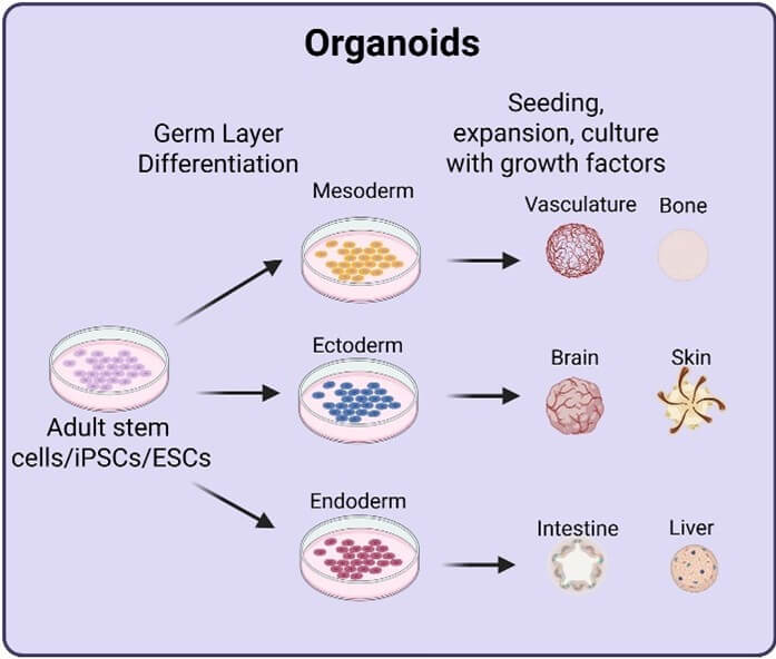
Organoids are three-dimensional multicellular structures grown in vitro that mimic key functional and structural aspects of real human organs. They are usually derived from embryonic stem cells or induced pluripotent stem cells, but can also come from adult stem or progenitor cells isolated from tissue biopsies.17 By providing the right developmental cues—growth factors, extracellular matrix components, or mechanical signals—scientists can guide stem cells to self-organize into organoids that exhibit the cell diversity, polarity, and basic functions of the intended human organ.3,17 Different types of organoids have been developed, including gastrointestinal, liver, brain, kidney, and lung organoids.18
Organoids are used to model human development and disease, and to evaluate drug response in a physiological and human-relevant context. Compared to experiments on animals, organoids provide human-specific insights, raise fewer ethical concerns, and deliver results faster (weeks rather than months or years).17 They also allow for high-throughput studies and personalized medicine, since organoids can be made from a patient’s own cells to predict responses to specific treatments.19 For example, patient-derived intestinal organoids have been used to test responses to cystic fibrosis therapies, accounting for each patient’s unique genetic and environmental history to aid in the development of tailored therapeutic strategies.17 Brain organoids are being used to study cerebral cortex development, neurodevelopmental disorders, and psychiatric conditions such as autism, schizophrenia, and bipolar disorder.7
Bioprinted organoids represent the next step. Here, bioinks made of cells, biomaterials, and growth factors are “printed” layer by layer into precise structures. This allows for more accurate arrangement of cells, biomaterials, growth factors, and other components. In addition, this technique enables greater control over shape and size, producing organoids that are even more physiologically relevant to real organs.19
Organs-on-chips

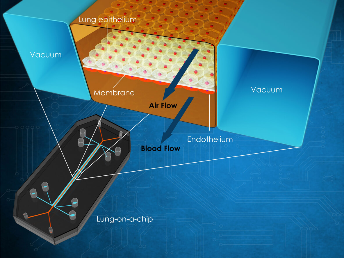
An organ-on-a-chip (OoC) is a microfluidic device that recreates aspects of an organ’s environment, dynamics, and physiological responses in real time.20 These chips are made of glass or polymers, with multiple channels separated by porous membranes where human cells are cultured. When connected to a peristaltic pump, the system allows for realistic nutrient exchange, mechanical forces such as breathing or blood flow, and cell-cell interactions.3,20 OoCs have been developed for the heart, kidney, lung, intestine, liver, and brain, as well as linked, multi-organ systems that model interactions between organs.3,21–23
Like organoids, OoCs are used to study human disease and drug response. Examples include a cardiac ischemia-on-a-chip to study hypoxia,21 an alveolus-on-a-chip to investigate SARS-CoV-2 infection,21 a vagina- and cervix-on-chip to model bacterial vaginosis and test potential treatments,7 and a lung-on-chip to gain insights into the airway during cystic fibrosis.22
OoCs share similar advantages over animal models as organoids: They’re human-specific, more ethical, higher throughput, and offer faster timelines and the potential for personalized medicine. They can also reproduce complex features of organs such as tissue interfaces, immune cell interactions, and physiological mechanical cues, including the dynamic fluid flow of an in vivo human organ. When combined, they can provide cellular and molecular insights into a disease that involves multiple tissue types.7 Organ chips have already shown how well they can replicate complex human responses to diseases. In addition to Emulate’s liver-on-a-chip, a lymphoid follicle chip was able to replicate responses to vaccines for influenza and SARS-CoV-2.7
Computational modeling
Advances in next-generation sequencing and computing have expanded the use of mathematical and computational models to simulate biological processes. These models allow scientists to test hypotheses, predict outcomes, and design new experiments. At the molecular level, systems models can be used to study biochemical processes, cell signaling, protein interactions, and gene regulation.24 Applications include digital twins, which are virtual representations of individuals that integrate clinical, genetic, molecular, environmental, and social data to predict disease progression and test treatment strategies.25 Recently, a multi-scale, multi-approach computational model was used to study the dynamics of T cells at the molecular, cellular, and systemic scales during influenza infection.24 Using a machine learning framework, scientists predicted the severity of F8 mutations in hemophilia A to guide personalized treatment strategies.26 Researchers have also identified potential colorectal cancer drug targets using a systems biology approach.27
Why haven’t NAMs been more widely adopted by the biomedical research field?
Despite the availability of numerous NAMs—many of which have shown great promise and even outperformed animal studies—they are still not as widely used in biomedical research as they should be. Workshops and surveys of biomedical researchers have helped clarify the reasons. Researchers have expressed uncertainty about their reliability, stemming from limited knowledge and a reluctance to adopt unfamiliar approaches.3,28 This hesitation persists, even though experiments on animals fail to predict whether a new drug will be safe and effective in 95% of clinical trials.29 There is a clear double standard: experiments on animals have been presumed to be the “gold standard,” yet they have never been rigorously standardized or validated to reliably determine how a drug will behave once in the human body. It’s good that NAMs are undergoing rigorous assessment to demonstrate reliability before widespread adoption, but animal-based methods were never held to this same standard. This is also why NAMs should be validated against human—not animal—data.
Additional barriers include concerns about whether NAM-only studies will be accepted for publication and in grant applications.3,30 In a survey of 68 researchers, about 31% reported that they preemptively conducted experiments on animals, assuming reviewers would request this data, while 44% said they had been explicitly asked by reviewers to add animal data.31 This highlights the presence of “animal methods bias” in scientific publishing, which discourages researchers from relying solely on NAMs for fear of rejection and career consequences. The impact of this bias extends beyond individual studies. When publication is less accessible for NAM-based research, early-career scientists may feel pressured to rely on experiments on animals to secure funding, tenure, or recognition. This creates a self-reinforcing cycle in which animal-based research dominates the scientific field, regardless of whether NAMs provide more relevant or predictive data for human health.
Funding is another major concern. In 2024, NIH launched Complement-ARIE, a program dedicated to the development, standardization, and validation of NAMs.28 While this represents an important step forward, the financial support allocated is disproportionately small. The proposed 2025 budget for Complement-ARIE was $35 million32—roughly 700 times less than what NIH typically spends annually on experiments on animals.33 Limited investment in NAMs restricts the ability to refine, validate, and scale up these methods. Meanwhile, experiments on animals benefit from entrenched infrastructure and decades of steady funding, giving them a substantial institutional advantage. Without a meaningful rebalancing of resources, the transition away from animal-based approaches will remain stalled—even though NAMs hold far greater promise for advancing human health.
How can this be solved?
To encourage wider adoption of NAMs, educational opportunities such as university courses, webinars, and conference workshops intended for researchers at all career stages could help build awareness and expertise. Some of these resources already exist, such as the European Commission’s Joint Research Centre Summer School on Non-Animal Approaches in Science34 and the Physicians Committee for Responsible Medicine’s Early-Career Researchers Advancing 21st Century Science program.35 However, these programs reach a limited audience. If NIH or U.S. universities offered similar initiatives on a larger scale, they could significantly broaden access, particularly for early-career scientists and researchers in institutions that lack NAM expertise. Expanding educational opportunities would also foster a research culture in which human-relevant, animal-free methods are seen as standard practice rather than niche alternatives.
Publishing practices must also evolve to address animal methods bias. The Coalition to Illuminate and Address Animal Methods Bias is actively working to mitigate this issue and has created an author guide to help researchers publish animal-free studies.36 Addressing this issue requires coordinated action at multiple levels, including training reviewers to recognize and fairly evaluate NAM-based research, journals enacting policies that explicitly accept high-quality non-animal studies and developing clear, standardized criteria for assessing NAM validity and rigor. Over time, such reforms could normalize NAMs in scientific literature and remove systemic barriers that currently discourage their adoption.
Increased funding opportunities for NAMs research would further accelerate both adoption and improvement of existing methods.28,30 NIH could address this by reallocating funds from animal experiments that have consistently failed to provide effective treatments toward NAM-focused initiatives. This could include increasing the Complement-ARIE budget or issuing funding calls exclusively for NAMs. Expanding financial support would not only enable more robust development of NAMs but also signal a broader institutional commitment to modernizing biomedical research practices.
Summary and conclusion
A wide range of NAMs already exists with the potential to advance disease research faster, more effectively, and more cost-efficiently compared to continued reliance on animal models. Human-relevant models—including 3D culture systems, multi-omics, and advanced computational approaches—offer insights that are often impossible to obtain from animal studies, particularly for complex human diseases. Continuing experiments on animals to study human diseases slows scientific progress, wastes resources, and misdirects research efforts due to poor translatability. While NIH has begun to recognize this, sustained momentum and strategic investment are necessary. By expanding educational initiatives, addressing publication biases, and increasing targeted funding, NIH and other government health agencies can promote broader adoption of NAMs, accelerate scientific breakthroughs, and ultimately improve human health outcomes.
- Trunnell ER. Breaking: Human-based research takes center stage at NIH. Science Advancement & Outreach. April 2025. Accessed September 18, 2025. https://www.scienceadvancement.org/reflections/human-based-research-takes-center-stage-at-nih/
- Trunnell ER. What happens when NIH stops asking for animal experiments? Science Advancement & Outreach. July 2025. Accessed September 18, 2025. https://www.scienceadvancement.org/reflections/what-happens-when-nih-stops-asking-for-animal-experiments/
- Hope L, Bailey J. Breaking down the barriers to animal-free research. Altern Lab Anim. 2025;53(4):215-231. doi:10.1177/02611929251349465
- Nahle Z. Making NAMs a ‘Complement’ to animal experimentation is the Trojan Horse of the animal-industrial complex—a scheme to nullify the principles of the 3Rs and void the quest to ‘replace’ animals for good! NAM J. 2025;1:100030. doi:10.1016/j.namjnl.2025.100030
- Wild D. Alternatives to animal testing: a tech run-down. In Vivo. January 6, 2025. Accessed September 18, 2025. https://insights.citeline.com/in-vivo/innovation/alternatives-to-animal-testing-a-tech-run-down-QSFFYKWSYBARTOLO5X3ZAVG62E/
- Reardon S. Beyond lab animals. Science. 2025;389(6761):676-679. doi:10.1126/science.aeb3933
- Taylor-Smith K. Bridging the gap: creating more physiologically relevant human cell models. Technology Networks. April 9, 2025. Accessed September 18, 2025. https://www.technologynetworks.com/cell-science/articles/bridging-the-gap-creating-more-physiologically-relevant-human-cell-models-396896
- PETA UK. Non-animal research methods and tests. Accessed September 18, 2025. https://www.peta.org.uk/issues/animals-not-experiment-on/non-animal-research-methods/
- Cruelty Free International. Alternatives to animal testing. Accessed September 18, 2025. https://crueltyfreeinternational.org/about-animal-testing/alternatives-animal-testing
- Cohen J. The last gift: how bodies donated for research may help find a cure for HIV. Science. September 4, 2025. Accessed September 18, 2025. https://www.science.org/content/article/last-gift-how-bodies-donated-research-may-help-find-cure-hiv
- Bledsoe MJ, Grizzle WE. The use of human tissues for research: what investigators need to know. Altern Lab Anim. 2022;50(4):265-274. doi:10.1177/02611929221107933
- Physicians Committee for Responsible Medicine. Access to human tissues. Accessed September 18, 2025. https://www.pcrm.org/ethical-science/animal-testing-and-alternatives/access-human-tissues
- Physicians Committee for Responsible Medicine. Human tissue research. Accessed September 18, 2025. https://www.pcrm.org/ethical-science/animal-testing-and-alternatives/human-tissue-research
- Chen C, Wang J, Pan D, et al. Applications of multi‐omics analysis in human diseases. MedComm. 2023;4(4):e315. doi:10.1002/mco2.315
- Harding P, Owen N, Eintracht J, et al. Variant-specific disruption to notch signalling in PAX6 microphthalmia and aniridia patient-derived hiPSC optic cup-like organoids. Biochim Biophys Acta BBA – Mol Basis Dis. 2025;1871(6):167869. doi:10.1016/j.bbadis.2025.167869
- Lindsay D, Adapa RM, Menon DK, Stamatakis EA. The amnesic effects of propofol on functional connectivity in the hippocampus determined by functional magnetic resonance imaging in volunteers. Br J Anaesth. Published online June 3, 2025. doi:10.1016/j.bja.2025.04.032
- Sidharthan C. Could organoids make animal models obsolete in research? News Medical. May 2, 2025. Accessed September 18, 2025. https://www.news-medical.net/health/Can-Organoids-Replace-Animal-Models-in-Human-Disease-Research.aspx
- Zhu Z, Cheng Y, Liu X, et al. Advances in the development and application of human organoids: techniques, applications, and future perspectives. Cell Transplant. 2025;34:09636897241303271. doi:10.1177/09636897241303271
- Li Z, Li K, Zhang C, et al. Bioprinted organoids: an innovative engine in biomedicine. Adv Sci. 2025;12(33):e07317. doi:10.1002/advs.202507317
- Dynamic 42. What is organ-on-chip? May 28, 2025. Accessed September 19, 2025. https://dynamic42.com/what-is-organ-on-chip/
- Dynamic 42. Organ-on-chip applications by organ type—what has been done? July 9, 2024. Accessed September 19, 2025. https://dynamic42.com/organ-on-chip-applications-by-organ-type/
- Ingber DE. Human organs-on-chips for disease modelling, drug development and personalized medicine. Nat Rev Genet. 2022;23(8):467-491. doi:10.1038/s41576-022-00466-9
- National Center for Advancing Translational Sciences. Meet chip. July 3, 2025. Accessed September 19, 2025. https://ncats.nih.gov/research/research-activities/tissue-chip/meet-chip
- Puniya BL, Verma M, Damiani C, Bakr S, Dräger A. Perspectives on computational modeling of biological systems and the significance of the SysMod community. Zhu S, ed. Bioinforma Adv. 2024;4(1):vbae090. doi:10.1093/bioadv/vbae090
- Katsoulakis E, Wang Q, Wu H, et al. Digital twins for health: a scoping review. Npj Digit Med. 2024;7(1):77. doi:10.1038/s41746-024-01073-0
- Lopes TJS, Rios R, Nogueira T, Mello RF. Prediction of hemophilia A severity using a small-input machine-learning framework. Npj Syst Biol Appl. 2021;7(1):22. doi:10.1038/s41540-021-00183-9
- Tavakoli N, Fong EJ, Coleman A, et al. Merging metabolic modeling and imaging for screening therapeutic targets in colorectal cancer. Npj Syst Biol Appl. 2025;11(1):12. doi:10.1038/s41540-025-00494-1
- Sunderic K, Wright AM, Kleinstreuer N, et al. Complement-ARIE: catalyzing the development and adoption of new approach methodologies. NAM J. 2025;1:100026. doi:10.1016/j.namjnl.2025.100026
- National Center for Advancing Translational Sciences. New therapeutic uses. November 5, 2024. Accessed September 22, 2025. https://ncats.nih.gov/research/research-activities/ntu
- Oyetade O, Allen DG, Carder J, et al. Ways to broaden the awareness, consideration and adoption of new approach methodologies (NAMs). ALTEX. Published online 2025. doi:10.14573/altex.2505281
- Krebs C. A survey to assess animal methods bias in scientific publishing. ALTEX. Published online 2023. doi:10.14573/altex.2210212
- Division of Program Coordination, Planning, and Strategic Initiatives. Complement Animal Research in Experimentation (Complement-ARIE) – A Common Fund Proposal. National Institutes of Health; 2024. Accessed September 23, 2025. https://dpcpsi.nih.gov/sites/default/files/2024-01/1-1PM-OSC-ConceptComplement-ARIE-Rutter-Woychik-onepager-508.pdf
- Institute of Medicine, National Research Council. Emerging Legal Trends Impacting Animal Research. In: International Animal Research Regulations: Impact on Neuroscience Research: Workshop Summary. National Academies Press (US); 2012. Accessed September 23, 2025. https://www.ncbi.nlm.nih.gov/books/NBK100123/
- Joint Research Centre (European Commission). EU Reference Laboratory for alternatives to animal testing (EURL ECVAM) – European Commission. 2025. Accessed September 23, 2025. https://joint-research-centre.ec.europa.eu/projects-and-activities/reference-and-measurement/european-union-reference-laboratories/eu-reference-laboratory-alternatives-animal-testing-eurl-ecvam_en
- Physicians Committee for Responsible Medicine. Engaging researchers in animal-free 21st century science. PCRM.org. 2025. Accessed September 23, 2025. https://www.pcrm.org/ethical-science/ethical-education-and-training/ERA21
- Krebs CE, Camp C, Constantino H, et al. Author guide for addressing Animal Methods Bias in publishing. Adv Sci. 2023;10(30):2303226. doi:10.1002/advs.202303226
